Pathology of Celiac Disease
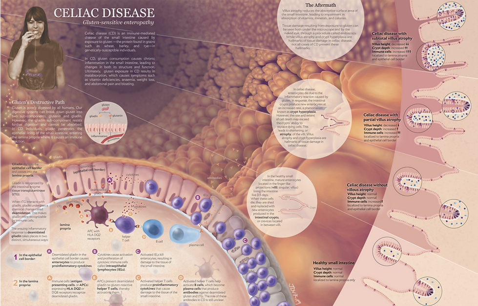
This double-page spread for a popular science magazine was created as part of the pathology course in the Biomedical Communications program. The goal of this piece is to communicate what is currently known about the etiology and pathogenesis of Celiac disease at the cellular, tissue, and organ/system levels. As disease presentation is heterogenous and its progression variable, a particular emphasis was placed on depiction of different pathological states of the affected tissue and organs, as opposed to changes in tissues and organs over time. Dr. John Wong, general pathologist and faculty member of the University of Toronto's Faculty of Medicine, served as the content adviser for this project, providing feedback on accuracy of both text and images.
Clients: Prof. Shelley Wall, Dr. John Wong, MD
Audience: educated lay audience
Format: print (magazine)
Media: Adobe Photoshop, Adobe Illustrator
Date: December 2018


Final illustration
Process work


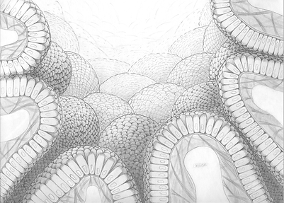
Preparatory Studies
The first stage of this project entailed a visual exploration of the effected tissues in celiac disease based on research conducted in the existing medical literature. The overarching goal of this exploration was to determine which visualization strategies might be most appropriate and effective for communicating about disease aetiology and pathogenesis.
A tissue landscape was created to better understand the visual 'landscape' in which the pathology occurs - i.e. the duodenum of the small intestine. Given the editorial nature of this project, I experimented with more creative visualization strategies.
Tissue cubes were created to gain an understanding of the possible variation in presentation of effected tissues. Due to the highly variable disease course observed amongst celiac disease patients, the focus of my cubes was discrete clinical presentations of the disease with no particular allusion to time. Given that tissue cubes are frequently used to illustrate changes over time, I recognized the possibility for misinterpretation in this regard when employing this visualization strategy. For this reason, it was determined that utilization of tissue cubes would not be appropriate for my final piece.

Advanced iteration

Intermediate iteration
Composition & Layout
Next, I began combining visual elements into a two-page story. The successes and challenges I encountered in my tissue studies guided my decisions regarding which visual representations to include.
Several iterations of the layout were produced in response to feedback from Dr. Wall and BMC peers. Initial presentation of visual elements was more geometric and grid-like; however, this clashed with the organic forms predominating in the scene (i.e. the plicae circularis, or folds of the small intestine). For this reason, the layout was modified to emulate the predominating organic forms.

Early iteration
Palette Selection
The final step before rendering was to determine a colour palette. Two main options were explored: automnal colours and jewel tones. The automnal palette was selected for its warm and cool colours, which more effectively communicate differences in healthy and disease states of tissue.

Automnal colours

Jewel tones
References
1. Adelman, D. C., Murray, J., Wu, T.-T., Mäki, M., Green, P. H., & Kelly, C. P. (2018). Measuring Change In Small Intestinal Histology In Patients With Celiac Disease. The American Journal of Gastroenterology, 113(3), 339–347. https://doi.org/10.1038/ajg.2017.480
2. Agur, A. M. R., & Dalley, A. F. (2009). Grant’s Atlas of Anatomy (12th ed.). Lippincott Williams & Wilkins.
3. Barker, J. M., & Liu, E. (2008). Celiac disease: pathophysiology, clinical manifestations, and associated autoimmune conditions. Advances in Pediatrics, 55, 349–65. https://doi.org/10.1016/J.YAPD.2008.07.001
4. Barret, M., Rahmi, G., Malamut, G., Samaha, E., & Cellier, C. (2015). Celiac Disease and Other Malabsorption States. In R. Kozarek & J. A. Leighton (Eds.), Endoscopy in Small Bowel Disorders (pp. 153–162). Switzerland: Springer International Publishing. https://doi.org/10.1007/978-3-319-14415-3
Chang, H. J., Burke, A. E., & Golub, R. M. (2011). Celiac Disease. JAMA, 306(14), 1614. https://doi.org/10.1001/jama.306.14.1614
5. Di Sabatino, A., & Corazza, G. R. (2009). Coeliac disease. Lancet (London, England), 373(9673), 1480–93. https://doi.org/10.1016/S0140-6736(09)60254-3
6. Fasano, A. (2009, August 1). Surprises from Celiac Disease. Scientific American, 301(2), 54–61. https://doi.org/10.1038/scientificamerican0809-54
7. Fasano, A., & Shea-Donohue, T. (2005). Mechanisms of Disease: the role of intestinal barrier function in the pathogenesis of gastrointestinal autoimmune diseases. Nature Clinical Practice Gastroenterology & Hepatology, 2(9), 416–422. https://doi.org/10.1038/ncpgasthep0259
8. Green, P. H. R., & Cellier, C. (2007). Celiac Disease. New England Journal of Medicine, 357(17), 1731–1743. https://doi.org/10.1056/NEJMra071600
9. Junqueira, L. C. U., Carneiro, J., & Kelley, R. (1996). Basic Histology (8th ed.). Appleton & Lange.
10. Kessel, R. G., & Kardon, R. H. (1979). Tissues and Organs: Text Atlas of Scanning Electron Microscopy. San Francisco: W.H. Freeman & Co Ltd.
11. Krstic, R. V. (1991). Human Microscopic Anatomy: An Atlas for Students of Medicine and Biology. New York: Springer-Verlag.
12. Kumar, V., Abbas, A. K., & Aster, J. C. (2018). Robbins Basic Pathology (10th ed.). Philadelphia, Pennsylvania: Elsevier.
13. Leonard, M. M., Sapone, A., Catassi, C., & Fasano, A. (2017). Celiac Disease and Nonceliac Gluten Sensitivity. JAMA, 318(7), 647–656. https://doi.org/10.1001/jama.2017.9730
14. Ovalle, W. K., & Nahirney, P. C. (2013). Netter’s Essential Histology (2nd ed.). Saunders.
15. Rubio-Tapia, A., Hill, I. D., Kelly, C. P., Calderwood, A. H., & Murray, J. A. (2013). ACG Clinical Guidelines: Diagnosis and Management of Celiac Disease. The American Journal of Gastroenterology, 108(5), 656–676. https://doi.org/10.1038/ajg.2013.79
16. Schuenke, M., Schulte, E., & Schumacher, U. (2010). Thieme Atlas of Anatomy: Neck and Internal Organs. New York: Thieme New York.
17. Stevens, A., Lowe, J. S., & Young, B. (2003). Wheater’s Basic Histopathology: A Color Atlas and Text (4th ed.). Churchill Livingstone.